This website uses cookies so that we can provide you with the best user experience possible. Cookie information is stored in your browser and performs functions such as recognising you when you return to our website and helping our team to understand which sections of the website you find most interesting and useful.
special hoof treatment &
orthopedic shoeing
Laminitis
Laminitis is one of the most painful orthopaedic diseases of the horse.
Laminitis is a diffuse aseptic inflammation of the hoof dermis (wall dermis). It is caused by interacting metabolic processes in the body. The pathological change on the hoof is only the local manifestation on the hoof. Usually both front hooves are affected.
Symptoms
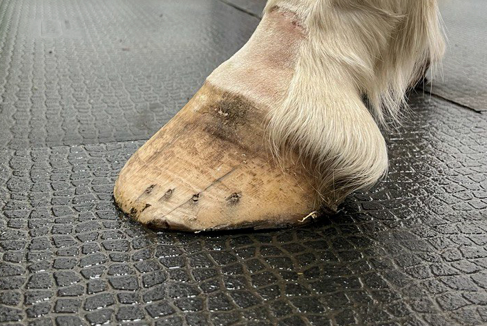
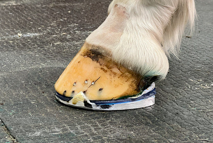
With the displacement of the coffin bone, the hoof can change in its external appearance.
A distinction is made between a knobby hoof and a knobless hoof. Reshaping hooves are only observed after long periods of disease. An optical change of the hoof crown occurs. In the acute or recurrent chronic stage, the cavities are filled with serofibrinous exudate due to the changes in the wall corium and the horn corium. The white line becomes very broad, fibrous and is partly bloodshot. The horn growth is increased in the toe area. A good farrier will provide orthopaedic shoeing about every five weeks.
Horses with chronic laminitis can often be recognised by the expert ( professional farrier, professional veterinarian) by the conspicuous heel-to-toe footing and the rapid flinging up of the hoof toe. Often a strong discolouration of the horn around the area of the frog tip is also observed. The horn of the sole is then coloured red from haemoglobin in this area.
Over the last 400 years, countless fittings have been developed. The best shoes are still those that relieve the hoof toe and raise the heels.
Therapy
The time of intervention is the measure of all things. It is best if medical help begins as early as 4 – 5 hours after the first clinical manifestations. First, of course, the feeding must be optimised. Only a small amount of hay is sufficient. The sodium / potassium balance must be optimised. Potassium deficiency leads to sodium excess – followed by high blood pressure and has a vasocontrictive (vasoconstricting) effect. Therefore, potassium chloride at 30g/day is recommended. Potassium chloride is available in pharmacies and can be administered orally. Biotin and zinc have a good growth effect for new hoof horn. If the horse allows it, the old shoes are removed. Plastic wedges are now glued under the hoof as soon as possible. Attaching them with hoof nails would only be cruelty to animals. The farrier must achieve immediate relief of the hoof toe and raise the hoof heights enormously. This will prevent the deep flexor tendon from tugging at the coffin bone. The bloodletting should be done by the vet and not the farrier. Our veterinarian has some effective medicines ready for acute laminitis.
Laminitis in the not yet chronic stage is curable; – if owner, farrier and veterinarian recognise and act early.
Chronic laminitis
Chronic laminitis begins when the coffin bone turns or sinks. If the coffin bone sinks by more than 10°, we speak of irreversible chronic laminitis (not curable). Already after 12 – 30 hours after the first clinical symptoms, the hoof dermis (the coffin bone carrier mechanism) can reject the coffin bone.
A minimum supply of oxygen and nutrients to keratogenic cells causes a loss of onychogenic substance and the formation of hoof horn of inferior quality. The suspensory apparatus of the coffin bone is observed in the toxic-chemical variant with destruction at the toe wall. In mechanical-traumatic laminitis, the destruction of the coffin bone carrier takes place on the sole.
thrush
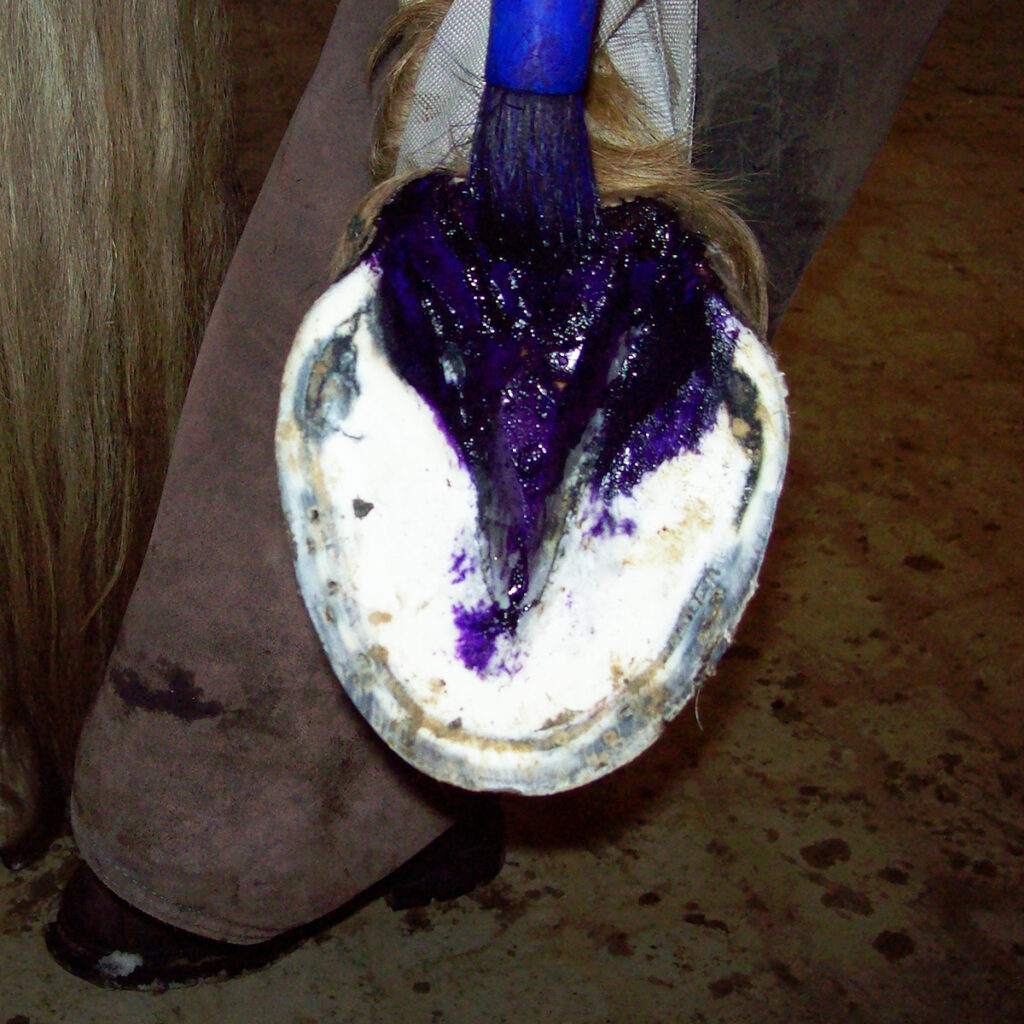
Thrush is a bacterial disease of the hoof. In this case, the soft horn of the frog is decomposed by putrefactive bacteria.
SYMPTOMS
- Putrefactive odour
- Cavities form in the frog, so-called pockets and cracks
- The firm tissue of the frog disintegrates into a greasy, crumbling mass
CAUSES
- Poor ground hygiene
- Poor hoof care
- Lack of exercise
- forced hoof
- contaminated soil
TREATMENT
- Eliminate causes
- Correct treatment of the hooves by a farrier.
- Drying substances, such as copper sulphate (undiluted or as a solution), potassium permanganate, iodoformether, should be applied after cleaning.
- Go to Equiwent and receive tailor-made help
Inflammations
coffin joint Inflammations
Coffin Joint inflammations are also divided into acute and chronic. Chronic joint inflammations generally lead to cartilage damage and bone formation (arthrosis).
Acute, infectious inflammation
Often caused by direct penetration of pathogens, e.g. through coronet steps. Spread of infections to neighbouring hoof organs.
The flexion test is not necessary in this case. Sometimes joint fistulas occur in cases that have spread.
Non-infectious
This is the most common form of inflammation, the sprain. “The horse has sprained its ankle”. Causes are always external influences on the joint capsule, ligaments or cartilage layer, e.g. Missteps, bruises, strains, twists, but also frequently incorrect trimming and poor shoeing
Chronical
If the hoof is constantly overloaded, the cartilage and the attachments of the joint capsule and the joint ligaments to the bones are constantly strained to a greater or lesser extent. Even if these strains do not lead to lameness, in the long run they cause ossification Cartilage damage becomes proliferation in the joint, up to stiffening.
Bone formation cannot be reversed. Therefore, in addition to eliminating the causes, treatment must focus on preventing further damage. Arthritic horses should be able to move as much as possible but calmly!
Special medication for cartilage nutrition and the
Correct trimming of the hoof and Orthopaedic shoeing is essential.
Often the front of the joint is damaged because the extensor tendon is directly attached to the edge of the capsule. Overstretching then hurts especially! This is another reason why rolling must be made easier. Protect the heels, toe direction! Sometimes it is necessary to use wedges or thickened shank ends.
navicular syndrom
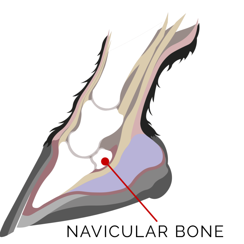
Navicular syndrome, navicular disease, and caudal heel pain are all referencing the same condition. Veterinarians have moved away from calling it navicular disease because disease means there is one problem, where syndrome means there are multiple or varying problems.
We now know that this condition has many components and it varies from horse to horse, so navicular syndrome is a more accurate description of what we are treating. It is often referenced as caudal heel pain as well to describe the location of the lameness; “caudal” meaning the back of the foot, and “heel pain” because that is the generalized area of the lameness.
Acute form
Irritation of the navicular bone area, especially the bursa under the deep flexor tendon.
Caused by overloading – this leads to the non-infectious form of navicular inflammation.
Injury, especially from nail treads.
Infectious form
The symptoms are similar to those of navicular fracture, but they are not as severe. In the infectious form, however, they are much more severe, and there is also fever, usually on the second day.
In the infectious form, there is a very high risk of the inflammation spreading to the hoof joint and the deep flexor tendon sheath, which can cause swellings extending beyond the fetlock joint!
Chronical form
This form has no infectious causes, so it is aseptic. It is one of the most significant causes of lameness in horses. A good knowledge of the anatomical situation is necessary to understand the causes and the changes that occur. When the foot is loaded, tensile and compressive forces occur at the navicular bone. At the points of application of these forces, wear and tear or changes in the bone and tendon structure as well as the bursa can occur.
The vet makes the diagnosis by x-raying and anaesthetising the responsible nerves on the usually lamer leg. If the other foot is also affected, the horse often goes lame on it after the injection!
The shoeing must of course ensure that the toes are short enough and the heels high enough. The latter is often achieved by wedges. This is acceptable, studs on the other hand are like poison. On softer ground they no longer have the desired effect, as they immediately sink down to the iron. The NBS (Natural Ballance Shoe) has gained a lot of importance in recent years.
Very important is the direction of the toes, which favours easy rolling of the hoof and thus helps to relieve the flexor tendons and navicular bone.
The veterinarians support the therapy with medications that promote blood circulation, which can achieve a lot today, provided that the hoof is always correctly trimmed.
Pododermatitis
Aseptic hoof dermatitis
Always caused by trauma, which is why it is usually called hoof bruising! Kicking, hitting, jumping on stone, high speed on hard ground . Continuous loading with less speed on hard ground tends to lead to ossification of the hoof cartilage. Poorly fitting horseshoes or poorly trimmed hoof.
Treatment:
- Sprue dressings
- Box rest with some leading on soft ground for about 3 – 4 days
- Then at least one week of restorative walking
- Orthopaedic shoeing
Septic hoof dermatitis
Caused by bacteria which can enter the hoof in many ways, e.g. nail tread or crown tread.
- Incorrectly cut stone pads
- Bacteria penetrating through horn gaps, etc.
In the case of very small inflammations - the horse will not go lame until pus (abscess) has formed
Treatment:
As for hoof abscesses, with additional antibiotics for larger processes.
Seedy Toe
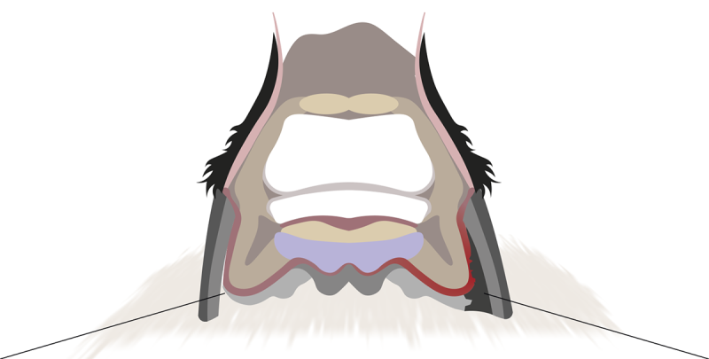
A seedy toe is a separation of the horn wall and the wall leather skin. It may extend upwards from the sole, i. e. the white line, to varying degrees.
Symptoms:
The white line is partially interrupted. In these areas, the horn substance is completely missing or weak and crumbly. When tapping (“percussion”) the wall areas in question, a hollow sound is obtained. Always compare with healthy areas!
Congenital causes
Wide hoof with sloping walls
The weight of the hoof on the bearing edge pushes the horn wall away from the corium at an angle (so-called shearing forces).
Poor horn quality
Pathological causes
Laminitis with (partial) detachment of the wall horn from the corium.
equine canker with destruction of the sole horn
Stroke with haematoma in the corium
rot in the area of the white line with dissolution of the horn
external causes
Incorrect hoof treatment
Poor hoof care and stable hygiene
Strong vertical rasping of the bearing wall horn with sloping hoof wall
The wall becomes too weak and breaks away to the outside or is slowly pushed away from the corium. Too narrow shoes or badly trimmed bearing rims. This leads to different pressure ratios between sole and bearing rim and thus to shearing forces.
With a loose wall, the risk of infection of the hoof corium is of course very high!
Treatment
Eliminate the cause.
Wide hooves have to be cut out especially often.
The wall horn is removed in the area of the defect. This is followed by disinfecting pressure bandages for two days. Then the defect is filled with putty. The bearing rim should remain suspended in this area until it heals. An orthopaedic shoe that supports the sole and relieves the weight-bearing rim of the hoof is recommended.
Wall seperation
A wall seperation is a contextual separation within the horn wall itself. Naturally, it sits at the transition between the tubular and lamellar layers of the horn. It usually originates from the sole.
Symptoms:
Splitting of the sole horn at the bearing edge.
Sometimes protrusion of the affected wall section
Sound change when tapped (percussion)
Lameness in severe cases
Causes:
Foreign body in the sole rim
incorrect nailing
crown fracture. In this case the defect starts from the top!
Hitting during jumping
Treatment:
Eliminate the cause
Let the defect grow out. Fill larger cavities with mastic. If the outer wall of the cavity is thin, it should be removed and replaced with putty. Again, shoeing is beneficial to support the wall and allow the defective bearing rim to float.
Bone spavin
Bone spavin is a gradual (chronic) bone disease of the hock joint. It is included in the list of hoof diseases because it is important for the farrier because of the therapy.
Like all arthrosis, bone spavin cannot be reversed and is therefore incurable. However, it is a form of arthrosis that can be treated relatively well and can therefore be “lived with” in many cases.
The treatment requires hoof orthopaedic as well as veterinary measures and a particularly good cooperation between farrier and veterinarian!
Trimming of the hoof
Relief of the inner joint parts. This means shortening the inner edges of the heels (smooth transition!) by approx. 2 – 3 mm. Orthopaedic shoeing is usually unavoidable.
Facilitate rolling: Protect the heels! Toe direction!
Veterinary measures
Medication to reduce inflammation and pain.
Surgery to cut the spat tendon and nerves in the critical joint area.
Surgery to stiffen the affected joint rows.
